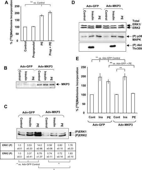
Figure 1. Overexpression of MKP3 blocks PE-induced activation of protein synthesis.
(A) Overnight cultures of cardiomyocytes were pretreated with 20 μM propranolol for 45 min before stimulation with 10 mM PE for 1.5 h. Protein synthesis was assayed as described in the Materials and methods section. Results are expressed as percentage change in [35S]methionine incorporation relative to the Adv-GFP control cells (±S.E.M.), which is set at 100%. Results are representative of three replicate experiments using separate preparations of ARVC, and each experiment comprised three replicates. S.E.M. is calculated from the averages of the three independent replicate experiments. **P<0.01 as indicated. (B–E) Freshly isolated ARVC were infected with Adv-GFP or Adv-MKP3 as indicated at an MOI of 100 pfu/cell. (B) Cells were treated with 10 nM insulin or 10 μM PE or DMSO (vehicle; control cells) for 5 min as indicated and then harvested. Lysates were analysed by SDS/PAGE/immunoblotting, as described in the Materials and methods section, and probed with anti-MKP3 antibody. (C) Cells were treated with 10 μM PE or 10 nM insulin for 5 min and harvested. Lysates were subjected to SDS/PAGE and immunoblots were analysed for phosphorylated ERK1/2 with anti-phospho-ERK1/2. Arrows indicate phosphorylated forms of ERK1 and ERK2. Densitometry was performed as described in the Materials and methods section, with ERK phospho-blots normalized to total ERK protein levels (determined by Western blotting, D) before comparison. Numbers are absolute values indicating fold change (±S.E.M.) in phosphorylated ERK1 and ERK2 relative to the Adv-GFP control sample. *P<0.05 as indicated. (D) Top panel: immunoblots were probed with an antibody for ERK1/2 to confirm equal loading. The middle and bottom panels show immunoblots that were analysed for phosphorylated p38 MAPK using anti-phospho-(Thr180/Tyr182) p38 MAPK or phosphorylated (Thr309) Akt antibodies respectively. Blots are representative of three independent replicate experiments using separate preparations of ARVC. (E) Cells were stimulated with PE (10 μM) or insulin (10 nM) for 1.5 h. Protein synthesis was assayed and results are presented as in (A).

