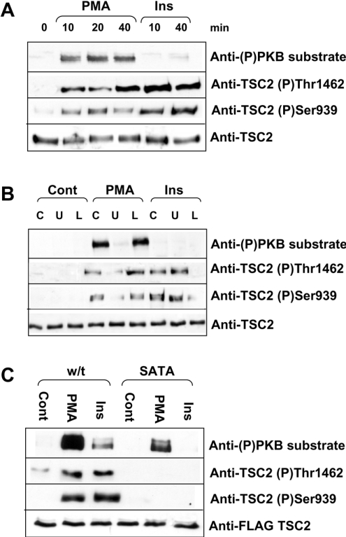
Figure 4. PMA induces the phosphorylation of TSC2.
(A–C) Immunoblots showing phosphorylation of TSC2 in response to PMA or insulin stimulation of HEK-293 cells. Topmost panels show TSC2 phosphorylated at serine/threonine residues within the sequence RXRXX(S/T). Second panels of (A–C) show TSC2 phosphorylated at Thr1462 and third panels show TSC2 phosphorylated at Ser939. The bottommost panels show total TSC2 as a loading control. (A) HEK-293 cells were treated with 1 μM PMA or 100 nM insulin for the time periods shown (min). (B) Cells were pretreated with 10 nM U0126 (U) or 30 μM LY294002 (L) for 45 min before stimulation with 1 mM PMA or 100 nM insulin for 20 min. (C) FLAG-tagged TSC1 and either wild-type (w/t) or SATA mutant TSC2 were overexpressed in HEK-293 cells. Cells were stimulated with 1 μM PMA or 100 nM insulin for 20 min. Cell lysates were harvested as described in the Materials and methods section. Endogenous TSC2 (A, B) or overexpressed, FLAG-tagged TSC1/2 (C) were immunoprecipitated from cell lysates and analysed by Western blotting as described in the Materials and methods section. Blots are representative of three independent experiments.

