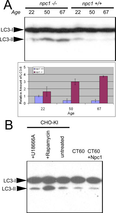
Figure 7. Levels of the Autophagosomal Marker LC3-II Are Increased in Degenerating npc1 Cerebellum.
(A) Immunoblots of 70 μg of protein resolved by 15% SDS-PAGE show increased levels of LC3-II in 50- and 67-d-old npc1 cerebellar extracts. Two mice of each age and genotype were analyzed, and relative band intensities were quantified. p-Values derived from comparing wild-type and npc1−/− cerebella at each age are 0.3, 0.01, and 0.001 for 22-, 50-, and 67-d-old mice, respectively.
(B) Immunoblots of CHO cell extracts resolved by 15% SDS-PAGE do not show increased levels of LC3-II in CT60 cells compared to wild-type CHO-KI cells or CT60 cells stably transfected with NPC1-YFP. No difference is noted with 10 μM U18666A, a drug that mimics npc1 loss of function. Rapamycin treatment (1 μM) of CHO-KI cells demonstrates robust activation of autophagy with increased LC3-II levels.

