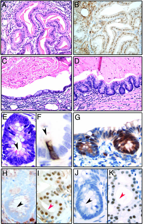
Fig. 2.
Analysis of p63–/– UGS grafts. (A) Hematoxylin and eosin stained section from a p63–/– graft showing glands lined by prostatic secretory cells (×200). (B) Prostatic luminal differentiation in p63–/– grafts is confirmed by Nkx3.1 immunoreactivity (×200). (C and D) Hematoxylin and eosin stained sections from a p63–/– graft showing mucous-producing cells (×200). These cells are either organized in a monolayer lining the glandular lumina (C) or form crypt-like structures that closely resemble colonic mucosa (D). The mucous cells in the crypts have the typical appearance of goblet cells. (E and F) High magnification of a crypt-like structure demonstrates the presence of Paneth cells, characterized by supranuclear eosinophilic secretory granules (E, black arrowhead, ×600) and positivity for lysozyme (F, black arrowhead, ×1,000). (G) Cdx2 is selectively expressed in the epithelial component of the p63–/– graft showing morphologic features of intestinal differentiation (×400X). (H–K) Nuclear immunoreactivity for Nkx3.1 (H and I) and AR (J and K) is absent in the mucinous epithelium showing features of intestinal differentiation (H and J, black arrowhead) and present in the prostate secretory cells (I and K, red arrowhead, ×600).

