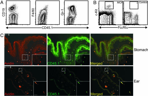
Fig. 2.
MCPs reconstitute mast cell compartments in mast cell-deficient KitW-sh/KitW-sh mice. Lethally irradiated KitW-sh/KitW-sh mice were injected i.v. with double-sorted CD45.1+ MCPs (104 cells per mouse) mixed with rescuing KitW-sh/KitW-sh bone marrow cells (3 × 10⁵ cells per mouse) or with rescuing KitW-sh/KitW-sh bone marrow cells only (negative control). Mice were killed 4 weeks after transplantation. (A) Flow cytometry analysis of the bone marrow from MCP-transplanted KitW-sh/KitW-sh mice. No donor-derived B cells (CD19), macrophages (F4/80), or granulocytes (Gr-1) were found. (B) Peritoneal lavage cells from MCP-transplanted or control KitW-sh/KitW-sh mice were analyzed by flow cytometry. Mast cells (gated population) were identified only in MCP-transplanted animals. (C) Immunofluorescence microscopy analysis of forestomach (Upper) and ear pinna (Lower) of MCP-transplanted KitW-sh/KitW-sh mice. Sections were stained with avidin for mast cells (red) and CD45.1 for donor-derived cells (green). Acquired images were then merged with photoshop (Adobe Systems, San Jose, CA). (Scale bars, 100 μm.) (Insets) Magnified views of the white boxed areas. Data shown are representative of those obtained in three experiments. (Scale bars, 10 μm.)

