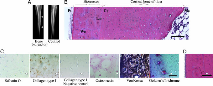
Fig. 2.
Histological characterization of the tissue within the bioreactor in absence of growth factors. (A) Radiograph of tibia with the bioreactor (right leg) and contralateral limb after 6 weeks. Arrowheads indicate top and bottom of bioreactor. (B) H&E-stained cross section of the bone bioreactor, adjacent cortical bone, and marrow cavity after 6 weeks. Ps, periosteum; bone, Wo, woven; Lm, lamellar; Ct, cortical; Ma, marrow. (Bar, 300 μm.) (C) Immunostaining for type I collagen and osteonectin and staining for hydroxyapatite mineral phase after 6 weeks. Arrowheads indicate demarcation between bioreactor space (on the left) and cortical bone (on the right). (Bar, 50 μm.) (D) H&E-stained cross section of the bone bioreactor and adjacent cortical bone after 8 weeks. Arrowhead indicates demarcation between bioreactor and tibia. (Bar, 250 μm.)

