Figure 2.
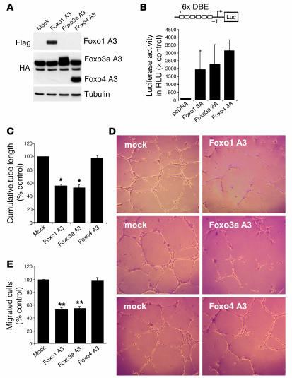
Overexpression of a gain-of-function mutant of Foxo1 or Foxo3a inhibits endothelial sprout formation and migration. (A) HUVECs were transfected with constitutively active Foxo1, Foxo3a, Foxo4, or mock control. Twenty-four hours later, cells were lysed and subjected to Western blot analysis with antibodies against Flag and HA. An antibody directed against tubulin was used as loading control. (B) HUVECs were transfected with a forkhead-responsive element reporter construct (6xDBE) along with plasmids encoding either constitutively active Foxo1, Foxo3a, or Foxo4. A transfected empty vector (pcDNA) was used a control. At 24 hours after transfection, cells were lysed, and luciferase relative to renilla luciferase activity was measured. × Control, fold value relative to pcDNA-transfected cells. The statistical summary represents the mean ± SEM; n = 3. (C and D) Statistical summary and representative micrographs of the tube-forming activity. HUVECs were seeded on Matrigel Basement Membrane Matrix 18 hours after transfection with the indicated plasmids. The length of capillary-like structures was measured by light microscopy after 24 hours in a blinded fashion. Data are presented as mean ± SEM; n = 5 (Foxo1), n = 6 (Foxo3a), n = 5 (Foxo4). *P < 0.001 versus control. Magnification, ×50. (E) HUVECs were transfected with the constitutively active constructs of Foxo1, Foxo3a, and Foxo4 and were seeded in the upper chamber of a modified Boyden chamber 18 hours after transfection. Endothelial cell migration was assessed using VEGF (50 ng/ml) as chemoattractant after 24 hours of incubation. Data are presented as mean ± SEM. **P < 0.05 versus control; n = 3.

