Figure 6.
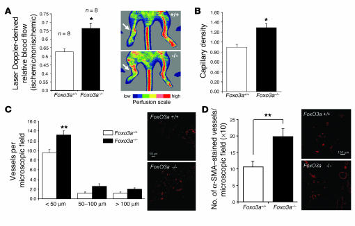
Foxo3a modulates neovascularization capacity in vivo. (A) Foxo3a+/+ and Foxo3a–/– mice were subjected to hind limb ischemia, and perfusion was assessed 14 days after onset of ischemia using laser Doppler imaging. Low or no perfusion is shown as dark blue, whereas the highest perfusion level is shown as red. Arrows indicate the ischemic leg. Quantitative results are presented as mean ± SEM; n = 8. *P = 0.002. (B) Capillary density (ratio of the number of capillaries to the number of myocytes) was determined in 8-μm frozen sections of the adductor and semimembraneous muscles. Quantitative results are presented as mean ± SEM; n = 8 (Foxo3a+/+), n = 7 (Foxo3a–/–). (C) Conductance vessels in the adductor and semimembraneous muscles were identified by size (>20 μm) and smooth muscle actin staining using a Cy3-labeled mouse monoclonal antibody for smooth muscle actin. The number of small (<50 μm), medium (50–100 μm), and large vessels was determined separately. Data are presented as mean ± SEM; n = 6 (Foxo3a+/+), n = 5 (Foxo3a–/–). **P = 0.01. (D) Statistical summary and representative micrographs of blood vessel infiltration in Matrigel sections stained with a smooth muscle actin antibody in wild-type and Foxo3a–/– mice. Quantitative results are presented as mean ± SEM; n = 7 (Foxo3a+/+), n = 8 (Foxo3a–/–). Scale bars in C and D, 100 μm.

