
Figure 2.
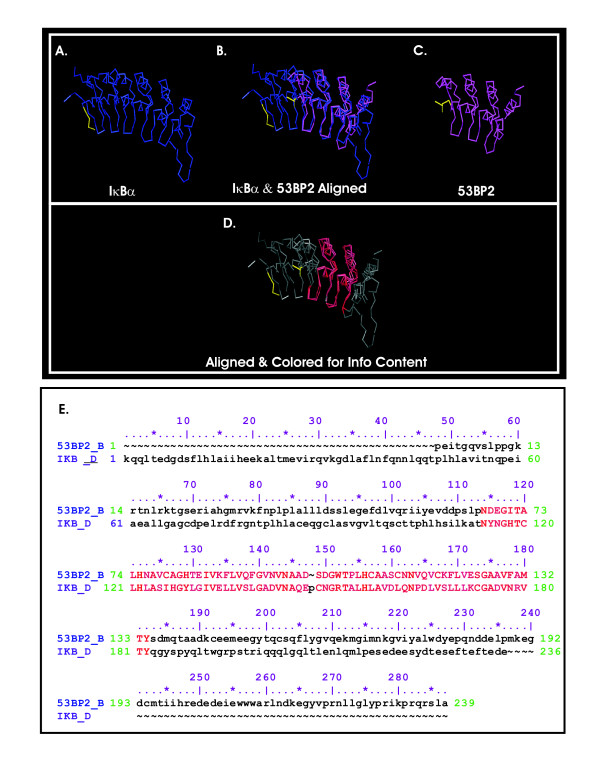
Alignment of IκBα (PDB ID:1IKN; 1IKN_D d1) with p53 binding protein 2 (PDB ID:1YCS; 1YCS_B d1). (A) The structure of IκBα (taken from the crystal structure of the p50/p65 heterodimer bound to IκBα); (B) The aligned structures of IκBα and 53BP2; (C) The structure of 53BP2 (taken from the crystal structure of p53 bound to 53BP2); (D) The aligned structure in 2B colored for conservation according to information content as described above; (E) The sequence alignment of IκBα and 53BP2 is depicted using the same coloring scheme as in the structure alignment in 2D. A four amino acid region near the N-termini of each structure is colored yellow as a reference point.