Figure 11.

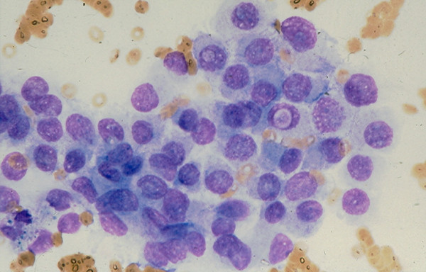
A loose sheet of tumor cells showing minimal nuclear crowding and two cells with intranuclear cytoplasmic inclusions in FNA of a conventional papillary carcinoma (Diff-Quik stain, × 400).

A loose sheet of tumor cells showing minimal nuclear crowding and two cells with intranuclear cytoplasmic inclusions in FNA of a conventional papillary carcinoma (Diff-Quik stain, × 400).