
Figure 22.
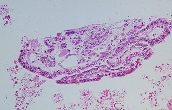
Papillary tissue fragments with thin fibrovascular cores covered with epithelial cells displaying nuclear crowding and occasional intranuclear cytoplasmic inclusions seen in a cell block section prepared from the needle aspirate of a papillary carcinoma with hemorrhagic cystic degenerative change (hematoxylin and eosin stain, × 250).