
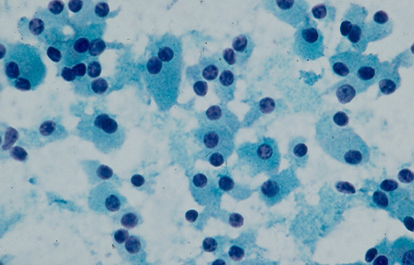
Figure 6.
Hurthle cells with abundant, granular cytoplasm and round, central or eccentrically located nuclei in FNA of a Hurthle cell lesion (Papanicolaou stain, × 400).
Official websites use .gov
A
.gov website belongs to an official
government organization in the United States.
Secure .gov websites use HTTPS
A lock (
) or https:// means you've safely
connected to the .gov website. Share sensitive
information only on official, secure websites.
Hurthle cells with abundant, granular cytoplasm and round, central or eccentrically located nuclei in FNA of a Hurthle cell lesion (Papanicolaou stain, × 400).