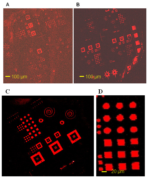
Figure 2.

Comparison between two types of slides. Fluorescence images of printed micronic patterns. Stamp was incubated with a 35-mers probe oligonucleotide for 30 sec, then put in contact for 15 sec with two types of microscope glass slides. A, electrostatic slide (ultra Gap, corning), B, dendrislide (home made slide). Slides were then incubated with a 15-mer 5'-Cy5 labeled oligonucleotide. C and D are a zoom area of B.
