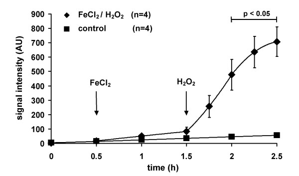
Figure 2.
The ESR signal intensity in isolated perfused and ventilatedrabbit lungs during baseline conditions and in the presence of FeCl2/H2O2. Lungs were either perfused with Krebs-Henseleit buffer containing 1 mM CPH only (control) or in the presence of FeCl2 (1 μM, added at 0.5 h, FeCl2/H2O2). After 1.5 h, H2O2 was added to the buffer fluid by continuous infusion (8 μmol/min) in the FeCl2-perfused lungs. Bar indicates significant differences between the FeCl2/H2O2 group and control.

