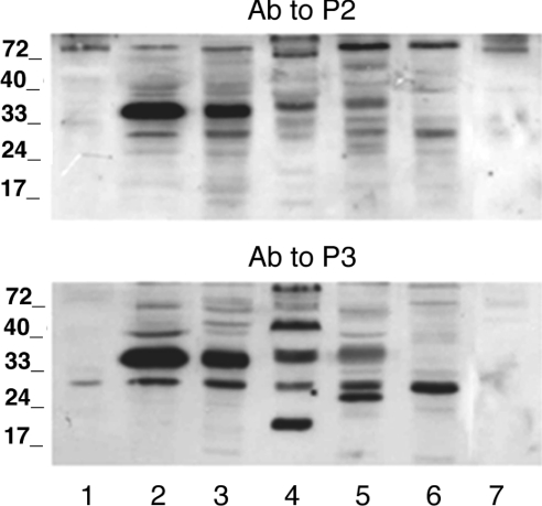
Figure 3. Immunoblot analysis of microsomes from human fibrocytes and different rat tissues.
Microsomal fractions were prepared as reported in the Experimental section. Microsomal proteins (20 μg) were separated by SDS/11% PAGE and blotted on to a nitrocellulose membrane. The blots were reacted with antibodies (Ab) against P2 (upper panel) or P3 (lower panel). Lanes 1, human fibrocyte; lanes 2, rat liver; lanes 3, rat kidney; lanes 4, rat skeletal muscle; lanes 5, rat brain; lanes 6, rat spleen; lanes 7, rat lung. The sizes of molecular mass markers (in kDa) are shown.

