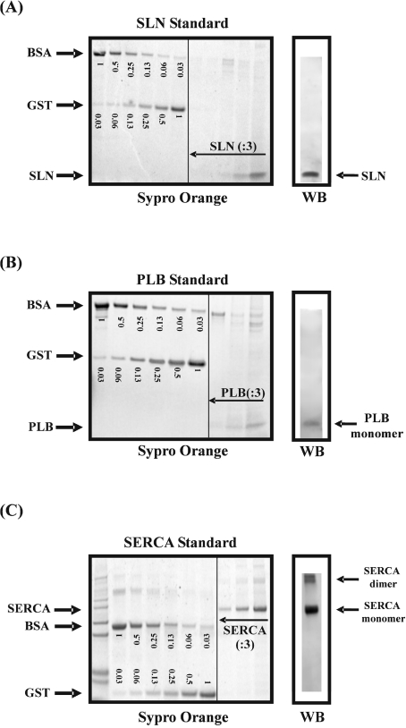
Figure 4. Purified SLN (A), PLB (B) and SERCA (C) proteins were evaluated by SDS/PAGE after staining with Sypro Orange.
In parallel, SLN, PLB and SERCA were identified via Western blotting (WB) using the SLNAP78, mA1 and TRY2 antibodies respectively. To determine the absolute amount of SLN, PLB and SERCA in the standard, the intensity of the purified protein bands on the Sypro-Orange-stained gel were compared with two protein standards of known concentrations: BSA and GST. On the gels, the amounts of BSA and GST are indicated in μg. Serial dilutions (:3) of SLN, PLB and SERCA were analysed.

