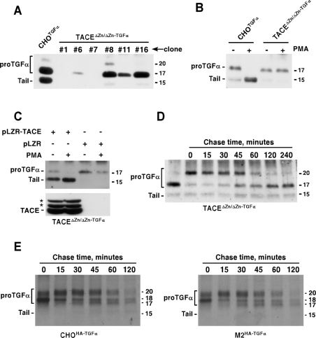
Figure 6. ProTGFα N-terminal shedding occurs in the absence of TACE activity.
(A) Expression of proTGFα in TACEΔZn/ΔZn cells. Cells were transfected with a vector coding for rat proTGFα, and clones selected. The expression of several clones with distinct levels of proTGFα is shown, compared with the expression in CHOTGFα cells. The immunoprecipitation and the Western blots were performed with the anti-proTGFα antibody. (B) Action of PKC activation by PMA on proTGFα cleavage. Where indicated, CHOTGFα or TACEΔZn/ΔZn−TGFα cells were treated with PMA (1 μM, 30 min), and the different proTGFα forms were analysed by Western blotting. (C) Reintroduction of TACE in TACEΔZn/ΔZn−TGFα cells rescues resting and PMA-induced cleavage. TACEΔZn/ΔZn−TGFα cells were infected with a retroviral vector containing TACE (pLZR-TACE), or with the empty retroviral vector (pLZR), and the expression of TACE was analysed by immunoprecipitation and Western blotting with an antibody that recognizes TACE (lower panel). The location of TACE is indicated, as well as two additional bands (asterisks) whose identity is unknown. The effect of reintroduced TACE on resting or PMA-induced (1 μM, 30 min) proTGFα cleavage was analysed by Western blotting (upper panel). (D) BB3103 causes reversible accumulation of N-terminal containing proTGFα forms in TACEΔZn/ΔZn−TGFα cells. TACEΔZn/ΔZn−TGFα cells were incubated overnight with 10 μM BB3103, and then chased in the absence of the drug for the indicated times. The different forms of proTGFα were analysed by Western blotting as above. (E) Wild-type or M2 mutant CHO cells expressing HA–proTGFα were labelled for 20 min with [35S]cysteine, and then chased for the indicated times in DMEM. Anti-HA immunoprecipitates were analysed by SDS/PAGE followed by autoradiography. Molecular masses are indicated in kDa in all panels.

