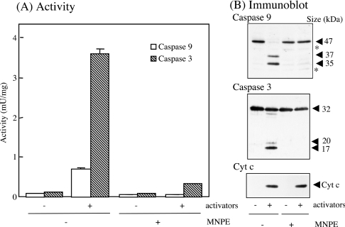
Figure 10. Effects of pre-treatment of cells with MNPE on caspase activation in the cell-free system.
HepG2 cells were pre-treated without (−) or with (+) 2 mM MNPE for 2 h, and cytosolic fractions were prepared immediately. Caspases in the fractions were activated by adding activators, cytochrome c, dATP and MgCl2. (A) Activities of caspase 9 and 3 were measured in triplicate. (B) Approx. 10 μg of protein was separated by SDS/PAGE and blotted on to the Hybond-P membrane. Immunoblot analyses were performed using the anti-(caspase 9), anti-(caspase 3) or anti-(cytochrome c) antibody. Sizes of proteins are shown in kDa. *, non-specific band. Typical data of triplicate experiments are shown.

