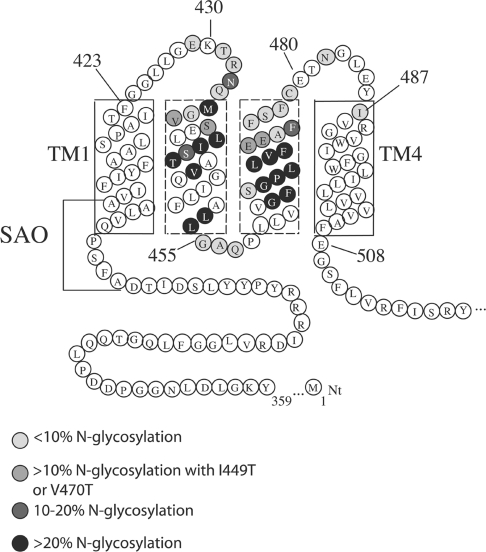
Figure 5. Refined model of the topology of TM1–4 in human AE1.
The first four TMs of human AE1 are shown, with residues involved in SAO deletion. The results of scanning N-glycosylation mapping are summarized. Position 429 cannot be N-glycosylated, as shown previously [20]. Residues are shaded according to their N-glycosylation efficiencies. Using the ‘12+14 rule’ places the lumenal end of TM1 at Phe423 and the beginning of TM4 at Ile487.

