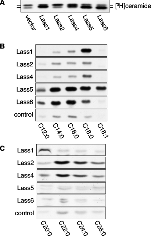
Figure 4. Comparison of ceramide synthesis activity of Lass family members.
(A) Lass proteins increase ceramide synthesis in cultured cells. HEK 293T cells transfected with plasmids as in Figure 3(A) were incubated for 4 h in medium with 1 μCi of [3H]dihydrosphingosine. Lipids were extracted and separated by TLC. (B) Comparison of in vitro ceramide synthase activities of Lass family members on fatty acyl-CoAs of various lengths and saturation. Samples (20 μg of protein) from the same cell lysates as used in Figure 3(A) were incubated with [3H]dihydrosphingosine/5 μM dihydrosphingosine and the indicated fatty acyl-CoA (25 μM) at 37 °C for 15 min. This experiment was performed three times with identical results. (C) Comparison of long-chain ceramide synthase activities of Lass proteins. Samples (40 μg of protein) from the cell lysates were incubated with [3H]dihydrosphingosine/5 μM dihydrosphingosine, the indicated fatty acyl-CoA (25 μM) and 0.1% digitonin (final concentration) at 37 °C for 15 min. The experiment was performed three times with identical results.

