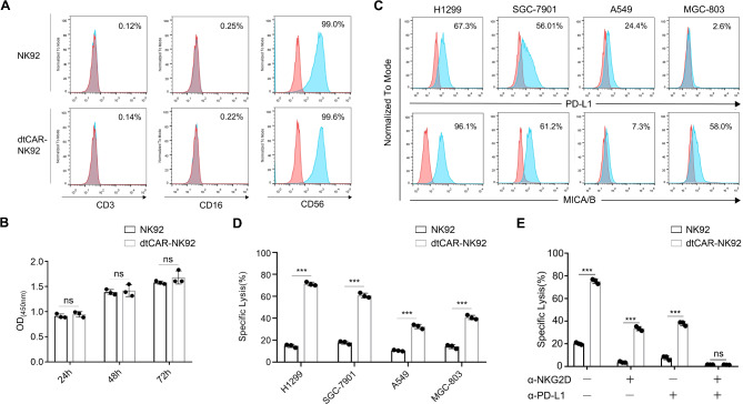
Fig. 2.
Phenotypic characterization and in vitro killing activity of dtCAR-NK92 cells. (A) The surface expression of phenotypic molecules on dtCAR-NK92 cells was analyzed by flow cytometry after staining with appropriate antibodies. (B) The proliferation of NK92 cells transduced with dtCAR was compared with parental NK92 cells using a CCK-8 assay. (C) Flow cytometry was used to analyze tumor cells stained with anti-PD-L1 and anti-MICA/MICB antibodies. (D) The cytotoxicity of dtCAR-NK92 cells was measured using the luciferase assay at effector-to-target ratios (E/T) of 5:1. (E) Assessment of the cytotoxicity of NKG2D- or PD-L1-blocked NK92 and dtCAR-NK92 cells against H1299 cells. Each experiment was performed with three independent replicates. Data are presented as the mean ± SD. Statistical analysis was performed using a two-tailed Student’s t-test; **p < 0.05, ***p < 0.001, ns: No significance

