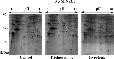
Figure 3. 2-D gel electrophoresis for protein extracts from the nuclei in which chromatin structural change, not DNA damage, was induced.
Protein extracts were prepared in 0.5-M NaCl salt solution as described in Figure 1, except for using the nuclei from HeLa cells that had been untreated (left-hand panel), treated with 10 μM TSA for 16 h (middle panel) or treated with hypotonic PBS containing 50 mM NaCl (right-hand panel) for 1 h. The extracts were separated on 2-D gels, as described in Figure 2. The intensity of the corresponding protein spots within the box, which was largely changed in Figure 2(C), was not changed by the treatment with TSA or hypotonic buffer.

