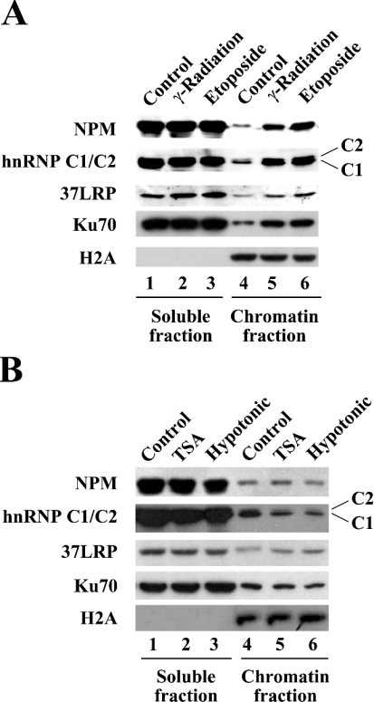
Figure 4. NPM and hnRNP C1/C2 bind to chromatin after DNA damage.
Chromatin-binding assays were performed. The soluble (lanes 1–3) and chromatin (lanes 4–6) fractions were separated using the nuclei from the cells in which DNA damage was induced as described in Figure 1(B) (A), or from the cells in which chromatin structural change was induced by treatment with TSA or hypotonic buffer (B). The distribution of the indicated proteins with soluble versus chromatin fractions was determined by immunoblotting with the antibodies indicated.

