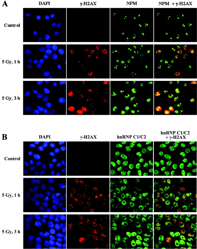
Figure 5. Examination of subcellular localization of NPM and hnRNP C1/C2 after DNA damage.
HeLa cells were exposed to 5 Gy of γ-radiation or treated with 50 μM etoposide (results not shown), and incubated for 1 or 3 h as indicated. The control cells were left untreated. Cells were then collected and subjected to dual immunostaining with antibodies against γ-H2AX (red) and NPM (green) (A), or γ-H2AX (red) and hnRNP C1/C2 (green) (B) before the immunofluorescence images were taken. The images of anti-γ-H2AX and anti-NPM staining, or anti-γ-H2AX and anti-(hnRNP C1/C2) staining were merged to examine co-localization (shown in the right-hand column in each row). The nuclei were visualized by DAPI staining (blue).

