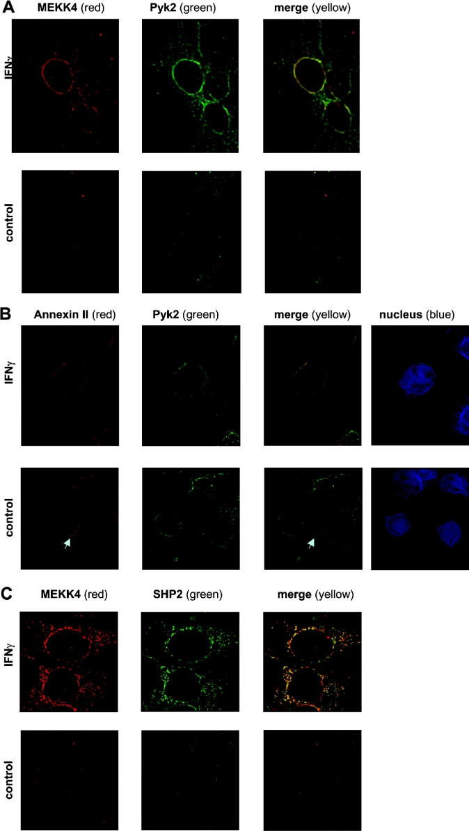
Figure 3. IFNγ-induced co-localization of MEKK4 and Pyk2, annexin II and Pyk2, and MEKK4 and SHP2 in the perinuclear region in HaCaT cells.
HaCaT cells, untreated or treated for 25 min with 200 units/ml IFNγ, were simultaneously stained with antibodies against MEKK4 and Pyk2 (A), annexin II (polyclonal) and Pyk2 (B), MEKK4 and SHP2 (C). The secondary antibody against MEKK4 (amino acids 18–139) and annexin II was Alexa-Fluor®-594-coupled anti-rabbit IgG, while Alexa-Fluor®-488-coupled anti-mouse was used against Pyk2 and SHP2 antibodies. Deconvoluted images of selected z-sections of immunofluorescence microscopy are shown, with MEKK4 and annexin II in red (first column) and Pyk2 and SHP2 in green (second column) and co-localization signals in yellow (third column). (B) Hoechst 33342 staining (blue) visualized the nucleus (fourth column). An arrow points to the accumulation of annexin II at points of cell–cell contact. Note that images in (A) are taken in a lower z-section showing more of the distribution in the cell body (closer to the cell-attachment area on the coverslip) than in (B), where most of the cell body was out of focus.

