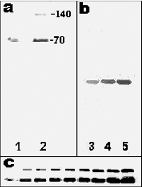
Figure 2. SDS/PAGE and Western blot analysis of the purified preparation (40 μg/ml).
(a) The inhibitor was run in denaturing conditions on a 12.5% polyacrylamide slab gel and revealed by silver staining. The volume of the preparation loaded in the wells were 1 μl (lane 1) and 3 μl (lane 2). (b) Inhibitor run in non-denaturing conditions and revealed by silver staining. The volume of the preparation loaded in the wells were 1 μl (lane 3), 3 μl (lane 4) and 5 μl (lane 5). (c) Analysis of the dimer formation by immunoblotting using the mEndopin 1 antibody. Sample volume loaded into the wells was from left to right: 1, 3, 5, 7, 9, 11, 15, 17 and 20 μl.

