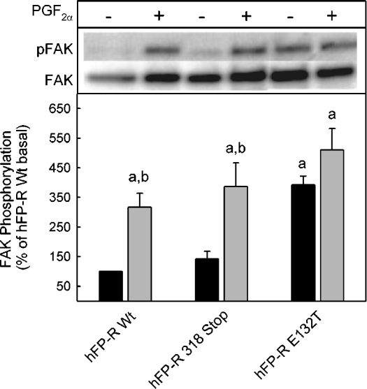
Figure 3. Constitutive activation of FAK in cells expressing the FP-E132T receptor.
Cells expressing wild-type FP-R or the indicated receptor mutant were incubated for 5 min in the presence or absence of 1 μM PGF2α, washed with PBS buffer and then lysed. The lysate was centrifuged and the supernatant was subjected to an immunoprecipitation with an FAK-specific antibody. Immunoprecipitated proteins were separated by SDS/PAGE and blotted on to PVDF membranes. Proteins were quantified with a phosphotyrosine-specific antibody (pFAK) and after stripping and restaining with an FAK-specific antibody, an appropriate peroxidase-labelled secondary antibody and a chemiluminescence substrate. Total (FAK) and phosphorylated (pFAK) immunoreactivities on the same blot are given. Values were normalized to the values of FAK and pFAK in the absence of PGF2α in cells expressing the wild-type (Wt) receptor. They are means±S.E.M. for three independent experiments. Statistics: Student's t test; a, significantly different from wild-type non-stimulated control; and b, significantly different from respective non-stimulated cells.

