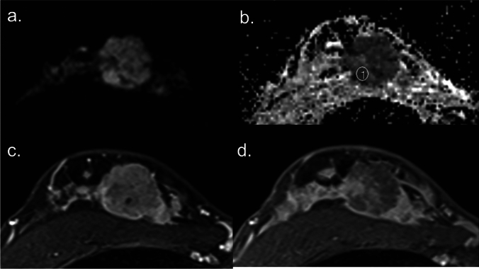
Fig. 1.
Diffusion-weighted (DW) image with b value = 1000 sec/mm2 (a), corresponding apparent diffusion coefficient (ADC) map (b), early phase dynamic contrast-enhanced (DCE) image (c), and delayed phase DCE image (d) of breast cancer. The breast cancer exhibits hindered diffusion and hyperintense on the DW image (a) with a correspondingly low ADC value of approximately 0.77 × 10⁻³ mm2/s on the ADC map (b). The lesion demonstrates fast in the early phase (c) and washout enhancement in the delayed phase (d), characteristic of a malignant breast tumor.

