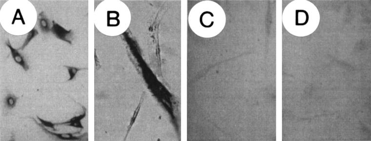
Figure 2.
Immunocytochemical localisation of insulin-like growth factor I (IGF-I) in non-transfected, parental, and in IGF-I antisense and triple helix transfected cells. Cells were stained for IGF-I by means of the immunoperoxidase technique using a polyclonal antibody against IGF-I. (A) parental (non-transfected) cells showing cytoplasmic staining for IGF-1; (B) cells transfected with a vector expressing β galactosidase showing cytoplasmic staining for X-gal; (C) cells transfected with pMT-anti-IGF-I; (D) cells transfected with pMT-AG triple helix. Note the change in morphology of antisense and triple helix transfected cells (C,D) compared with parental cells (A); the transfected cells do not express IGF-I .

