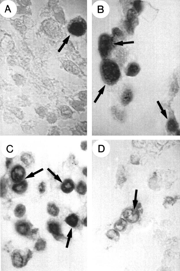
Figure 5.
Apoptosis detection in human glioma cells using the dUTP fluorescein terminal transferase labelling of nicked DNA technique (characteristic examples are marked by arrows). (A) Non-transfected cells; (B) insulin-like growth factor type I (IGF-I) antisense transfected cells; (C) IGF-I triple helix transfected cells; (D) IGF-I triple helix cells cotransfected with vectors encoding major histocompatibility complex class I (MHC-I) antisense and B7 antisense cDNA. Note the incorporation of labelled dUTP (dark staining with DAPI and fluorescein isothiocyanate) in apoptotic cells showing chromatin fragmentation: these cells are very rare in non-transfected cells (A), but are numerous in IGF-I antisense and triple helix transfected cells (B,C). On the contrary, apoptosis is infrequent in IGF-I triple helix cotransfected cells (D).

