Figure 1.
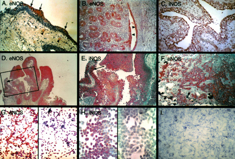
Nitric oxide synthase (NOS) in bladder cancer. (A) Normal bladder mucosa. Immunoreactivity for endothelial NOS (eNOS) is confined to the umbrella cells comprising the superficial layer (arrows). Positive staining is also noted in capillaries in the submucosa (arrowheads). (B) eNOS immunoreactivity in the fetal urothelium. Diffuse positive staining is seen in the urothelium of the renal pelvis (arrowheads) and in the epithelium of the collecting tubules (on the left side of the picture). (C) Diffuse inducible NOS (iNOS) immunoreactivity of the fetal bladder urothelium. (D) Immunoreactivity for eNOS in papillary transitional cell carcinoma of the urinary bladder. Most of the cells are positively stained. (E) Positive immunoreactivity for iNOS in papillary transitional cell carcinoma of the urinary bladder. Compare with negative eNOS immunoreactivity in the inset in D, which represents the same area (arrows). (F) Prominent immunoreactivity for eNOS in invasive bladder cancer induced by schistosomal infection. Degenerated eggs of the parasite are seen in the bladder wall (short arrows). (G) eNOS immunoreactivity in the UM-UC-3 bladder carcinoma cell line. The negative control is on the right side. (H) iNOS immunoreactivity in the UM-UC-3 bladder carcinoma cell line. The negative control is on the left side. (I) Positive histochemical reaction with NADPH diaphorase demonstrating enzymatic activity of NOS (EJ28 bladder carcinoma cell line). The reaction gives a blue product in the absence of background staining.

