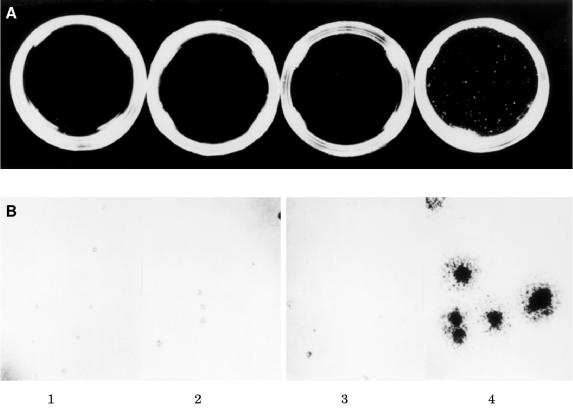
Figure 6.
Soft agar colonisation of EMT6 murine mammary carcinoma cells. Assays were performed in parallel on all cells under identical culture conditions. (A) Photographs are from a soft agar assay carried out on cells from the H, J, and O clones (panels 1, 2, and 3, respectively), carrying the antisense insulin-like growth factor I receptor (IGF-IR) construct and cells carrying the cytomegalovirus construct lacking the antisense IGF-IR insert (panel 4). Original magnification, ×1. (B) High power photographs of sample fields taken from assays depicted above. Original magnification, ×40.

