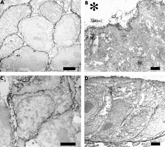
Figure 2.
Immunoelectron micrograph of regulatory proteins. (A) Membrane cofactor protein (CD46) in undifferentiated (poorly differentiated) adenocarcinoma. Unlike differentiated adenocarcinoma, the positive reaction is seen over the entire surface (bar, 1 μm; original magnification, ×4400). (B) Decay accelerating factor (DAF; CD55) in differentiated (papillary) adenocarcinoma. Note the positive reaction only on the luminal cell surface; N indicates the nucleus (bar, 1 μm; original magnification, ×3000). (C) DAF (CD55) in undifferentiated (poorly differentiated) adenocarcinoma. Unlike differentiated adenocarcinoma, the positive reaction is found over the entire surface (bar, 2 μm; original magnification, ×6000). (D) Protectin (CD59) in differentiated (tubular) adenocarcinoma. The strong reaction is found on the luminal and basal membrane, and the weak reaction on the lateral membrane (bar, 1 μm; original magnification, ×3000). The asterisk indicates the lumen of the cancer cells.

