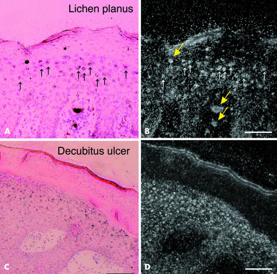
Figure 3.
In situ hybridisation for KLK8 mRNA in pathological skin. In situ hybridisation of KLK8 mRNA in the lesional skin of (A and B) lichen planus and (C and D) marginal skin around a decubitus ulcer. (A) And (C) are bright field micrographs of skin sections counterstained with haematoxylin and eosin; (B) and (D) are dark field micrographs of the same fields shown in (A) and (B), respectively. Silver grains were observed under light and dark field illumination (A–D). Small arrows in (A) and (B) represent cells labelled by the hybridisation probe. Thick arrows (yellow) represent artifacts (D). Scale bars: (A,B), 100 μm; (C,D), 200 μm.

