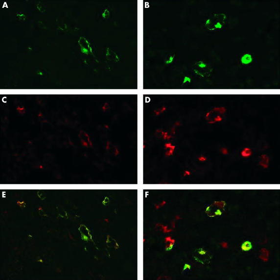
Figure 2.
Immunofluorescence staining for (A, B) latent membrane protein 1 (LMP1; green) and for (C, D) tumour necrosis factor receptor associated factor (TRAF1; red) shows similar patterns of labelling and overlaying the images reveals colocalisation of the signals (yellow) in most cells in (A, C, E) infectious mononucleosis and (B, D, F) an Epstein-Barr virus positive case of Hodgkin lymphoma. Some additional red staining is probably caused by the expression of TRAF1 in interdigitating reticulum cells.

