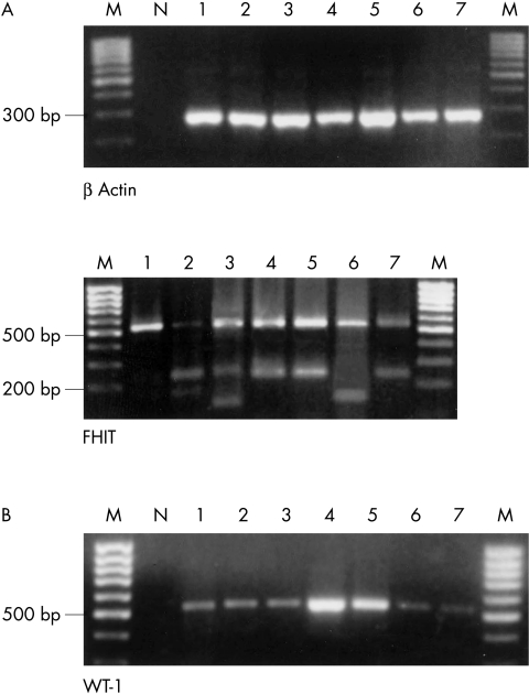
Figure 1.
(A) Agarose gel electrophoresis showing representative polymerase chain reaction (PCR) products of β actin reverse transcription PCR to demonstrate the integrity of the RNA (top panel). The analysis of FHIT (bottom panel) demonstrated in each case the full length product of 533 bp (lane 1) and in six cases various additional aberrant FHIT transcripts (lanes 2–7). The number of the lane corresponds to the histological diagnosis shown in table 2 ▶. (B) WT-1 mRNA was detectable in two benign and five malignant tumours (lanes 1–7). M, molecular weight marker; N, negative control.

