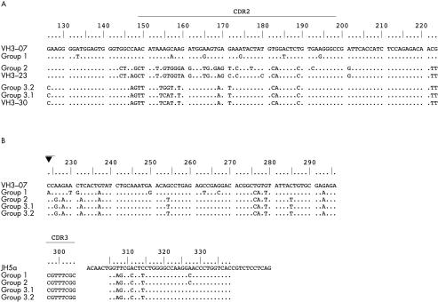
Figure 1.
Comparison of the immunoglobulin heavy chain (IgH) rearrangements of case 1 and alignment to the corresponding germline VH segments. (A) Alignment of the revised 5′ portions of the rearranged VH segments and the corresponding VH germline segments. Differences between the revised VH rearrangements, their most homologous germline segments, and the initial VH3–07 rearrangement, respectively, are shown in capital letters. This comparison demonstrates that these 5′-VH segments derive from different VH germline segments as a consequence of the VH receptor revision in this case. (B) Alignment of the common 3′ portions of the rearranged IgH genes to the corresponding VH germline segment (VH3–07) and JH germline segment (JH5a). All subclones of this case display the highest homology to the initial VH germline segment VH3–07, although the high mutation rate might mimic another germline segment. In addition, there are common and different somatic mutations, indicative of an ongoing somatic mutation process. These results prove that all subclones of this case carry the same 3′-VH rearrangement, which was not affected by the receptor revision. The closed triangle indicates the probable breakpoint for hybrid formation, just behind the last region of homology of the 5′-VH regions.

