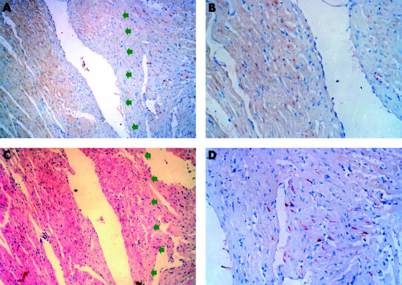
Figure 3.
Double staining (immunostaining for Fas and in situ terminal deoxynucleotidyl transferase mediated nick end labelling (TUNEL)) shows that Fas and TUNEL positivity were located in non-ischaemic myocardium without apoptosis and ischaemic myocardium, respectively. (A, C) Serial sections from a sample after 36 hours of ischaemia. (A) Double staining (red for Fas, brown for TUNEL). Note that Fas positive cells and TUNEL positive cells are distinct populations. Arrows point to the ischaemic region. (B) Magnification of a region positive by TUNEL. (D) Magnification of a region positive for Fas immunostaining. (C) Haematoxylin and eosin stain.

