Figure 1.
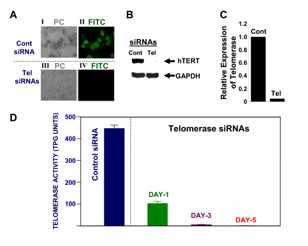
Effect of telomerase-specific siRNAs on telomerase expression and activity in SEG-1 cells. (A). Control (Cont) or telomerase specific (Tel) SiRNAs were transfected into Barrett's associated adenocarcinoma cells (SEG-1) and the cells were analyzed for protein levels of telomerase at day 7 by immunostaining, using a rabbit polyclonal antibody against telomerase. Antigen-antibody complex was located by incubation of cells with FITC-labeled anti-rabbit secondary antibody. Transfected cells within the same microscopic field were viewed and photographed by phase contrast (PC) or by fluorescence emitted at 518 nm (FITC filter). Using the FITC filter, telomerase positive cells appear bright green. (B). SEG-1 cells were treated exactly as described for panel "A" and telomerase expression was monitored by western blot analysis using a rabbit polyclonal anti-telomerase antibody. (C). Bar graph showing relative expression of telomerase in control (Cont) or telomerase (Tel) siRNA treated SEG-1 cells, as assessed by western blot analysis (shown in panel B). (D). SEG-1 cells were transfected with control (Cont) or telomerase specific (Tel) SiRNAs and evaluated for telomerase activity at days 1, 3, and 5 after transfection. Telomerase activity was evaluated using TRAPEZER XL Telomerase Detection Kit, a highly sensitive, quantitative, and non-isotopic version of the original Telomeric Repeat Amplification Protocol. Briefly the cell lysates (1000 cell-equivalents) were mixed with TRAPEZER XL reaction mix containing Amplifuor™ primers, incubated for 30 min at 30°C, and telomerase products generated by PCR were quantitated using a Fluorescence Plate Reader. Telomerase activity in SEG-1 cells treated with control siRNAs for 5 days and the cells treated with telomerase specific siRNAs for days 1, 3, and 5, is shown in TPG (total product generated) units.

