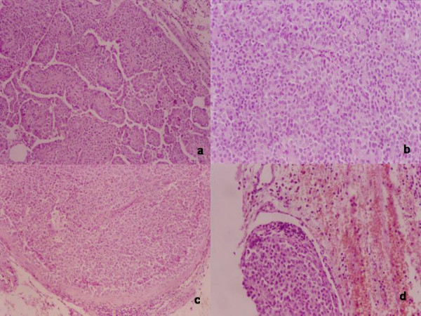
Figure 3.

Histological findings: H/E a) papillary growth pattern, focal nuclear pleomorphism and multinucleate giant cells; b) diffuse, sheet like, growth pattern c) massive infiltration in an encapsulated nodule, suggesting metastatic lymphnode; d) vascular invasion in the soft tissue surrounding the parathyroid;
