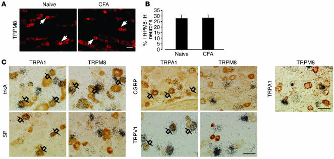
Figure 3.
No change of TRPM8 protein in the DRG and no overlap between TRPM8- and TRPA1-expressing neurons after inflammation. (A) Protein expression of TRPM8 in the naive DRG and the ipsilateral DRG at day 3 after inflammation, as detected by immunohistochemistry. TRPM8-immunoreactive (TRPM8-IR) neurons were invariably small or medium in size (arrows). (B) Quantification of the percentage of TRPM8-IR neurons at day 3 after CFA injection. (C) Double labeling by a combined method of ISHH and immunohistochemistry for TRPA1 or TRPM8 mRNA and trkA-IR, SP-IR, CGRP-IR, and TRPV1-IR in the DRG at day 3 after CFA injection. Double labeling for TRPA1 and TRPM8 mRNA by dual ISHH is shown at right. TRPA1- and TRPM8-expressing neurons were clearly distinguishable. Open arrows indicate double-labeled neurons. Scale bars: 50 μm.

