Figure 5.
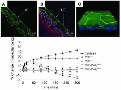
Localization of P2X3 in the uroepithelium and responses to pressure changes in P2X2- and P2X3-knockout mice. (A and B) Localization of P2X3 in cryosections of rabbit bladder uroepithelium. (A) P2X3 staining is shown in green, and the umbrella cells (UC) are marked with arrows. (B) Composite image with P2X3 staining shown in green, rhodamine-phalloidin–labeled actin in red, and Topro-3–labeled (Molecular Probes) nuclei in blue. (C) Whole mounted rabbit uroepithelium showing the distribution of the nuclei (blue) and P2X3 (green). The image is a 3-dimensional reconstruction of a Z series collected with a confocal microscope. The image was tilted around the x axis to emphasize the 3-dimensional aspect of the image. The grid is a 3-dimensional scale bar, with each side of the square approximately equivalent to 12.5 μm. (D) Bladders from mice of the indicated strains were mounted in Ussing stretch chambers, the pressure was increased at t = 0, and the capacitance was recorded. Data shown are mean ± SEM (n ≥ 3). *Statistically significant difference (P < 0.05) relative to the appropriate control.

