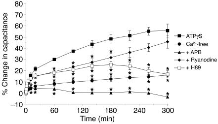
Figure 7.
Potential role of Ca2+ and PKA in ATPγS-stimulated changes in membrane capacitance. Rabbit uroepithelium was mounted in modified Ussing chambers and preincubated with 75 μM 2-aminoethoxydiphenylborate (APB), 50 μM ryanodine, or 10 μM H89 for 30 minutes as indicated. In the Ca2+-free experiments, the normal Krebs solution was isovolumetrically replaced with Krebs solution lacking Ca2+. At t = 0, 50 μM ATPγS was added into the serosal hemichamber, and changes in capacitance were monitored over time in the continued presence of the appropriate drug. Data are mean ± SEM (n ≥ 5). *Statistically significant difference (P < 0.05) relative to the ATPγS reaction.

