Figure 3.

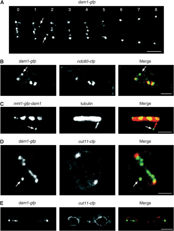
Dam1 binds non-kinetochore-associated spindle microtubule tips in mitosis. (A) Images from a movie of dam1-gfp cells in mitosis taken at 60 s intervals. Scale bar=2 μm. (B) High-magnification image of fixed dam1-gfp ndc80-cfp cells in mitosis. The arrow indicates non-kinetochore-associated Dam1. Scale bar=1 μm. (C) Log phase nmt1-GFP-dam1 cells were grown in the absence of thiamine for 24 h and fixed and stained with anti-tubulin antibody (tubulin). Scale bar=1 μm. The arrow shows Dam1 (green) on the plus end of an intranuclear spindle microtubule (red). (D) High-magnification image of dam1-gfp cut11-cfp cells before anaphase onset. Six Dam1 dots are observed in a line between the spindle poles. The arrow indicates a seventh Dam1 dot (green), which is inside the nuclear envelope (red). Scale bar=1 μm. (E) Image of dam1-gfp cut11-cfp cells after anaphase. Scale bar=3 μm.
