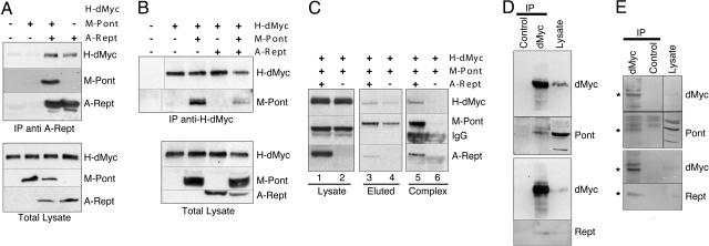
Fig. 1.
Rept and Pont associate with dMyc in vivo. (A and B) M-Pont and/or A-Rept were transiently transfected into S2 cells stably expressing H-dMyc (as indicated). (Upper) Whole-cell lysates were immunoprecipitated with anti-AU1 antibodies for A-Rept (A) or anti-HA antibodies recognizing H-dMyc (B), followed by immunoblotting with anti-tag antibodies to detect the proteins indicated on the right. (Lower) Immunoblots of whole-cell lysates to reveal the relative expression levels of the indicated proteins. Positions of H-dMyc, M-Pont, and A-Rept, respectively, are indicated. The first lane in B Upper contains lysate of nontransfected S2 cells. (C) M-Pont and A-Rept (lanes 1, 3, and 5) or M-Pont alone (lanes 2, 4, and 6) were transiently transfected into S2 cells stably expressing H-dMyc. Cell lysates were incubated with 9E10 antibodies, and the immunoprecipitate was eluted with the 9E10 peptide (lanes 3 and 4). The eluate was then reimmunoprecipitated with anti-AU1 antibodies (lane 5 and 6), and the immunoprecipitate was analyzed by immunoblotting. (D and E) S2 cell lysates (D) or third-instar larval extracts (E) were incubated with anti-dMyc antibodies or control hybridoma supernatant. Immunoprecipitates were blotted with anti-dMyc antibodies, anti-Pont, or anti-Rept antisera as indicated. The rightmost lanes show immunoblots of whole-cell lysates. Asterisks indicate the migration of the endogenous proteins.

