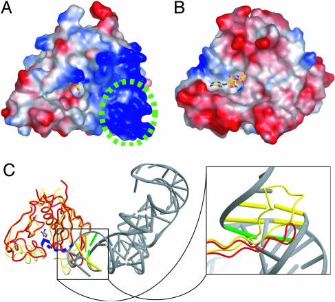
Fig. 6.
Recognition of tRNA moiety by AlaX. Shown is a surface potential representation of PhoAlaX (A) and ThrRS-N2 (8) (B), where acidic and basic potentials are represented in red and blue, respectively. The serine and the SerA76 are shown as stick models. The region corresponding to the hairpin motif is circled with a green dashed line. (C) Superposition of PhoAlaX (red tube) on ThrRS-N2–tRNA (yellow tube). (Left) The overview of the superposed image. The third base pair is shown as a green bar. (Right) An enlarged view of the hairpin motif interacting with tRNA.

