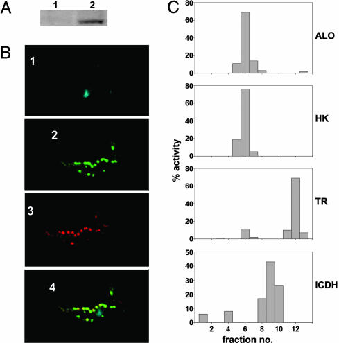
Fig. 6.
Localization of ALO to the glycosome of T. brucei and T. cruzi. (A) Blot containing lysates from 1 × 10⁷ T. brucei PCF cells expressing epitope-tagged TbALO from uninduced (lane 1) and tetracycline-induced (lane 2) cultures probed with a c-myc (9E10) monoclonal antibody. Protein loading was judged by Coomassie staining. (B) Tetracycline-induced T. brucei PCF cells expressing epitope-tagged TbALO were stained with DAPI (blue; 1), anti-9E10 (green; 2), and antiglycosomal GAPDH (red; 3). When the pattern of TbALO and glycosomal GAPDH staining was superimposed, colocalization was observed (yellow; 4). (C) T. cruzi epimastigotes (1 × 10¹⁰) were homogenized by using silicon carbide and fractionated on a continuous sucrose gradient (0.4–2 M). Fractions (0.75 ml) were collected and assayed for ALO, hexokinase (HK), trypanothione reductase (TR), and isocitrate dehydrogenase (ICDH) activity (Materials and Methods). The percent activity collected in each sample was determined.

