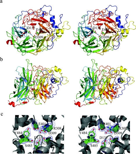
Figure 1. Three-dimensional structure of LsdA.
Superior (a) and lateral (b) stereo views of the five-bladed β-propeller fold. The colour is ‘ramped’ from N- (blue) to C- (red) terminus. Catalytic residues Asp135, Asp309 and Glu401 are shown in ball-and-stick representation. (c) Stereo view of the electron density map (contoured at 1σ level) ‘carved’ around catalytic residues and other residues involved in the hydrogen-bond (broken lines) network at the active site. These Figures were prepared with PYMOL [47].

