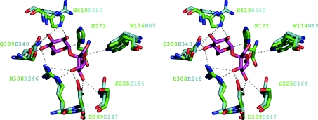
Figure 4. Sucrose-binding site of LsdA.
The Figure shows an overlap of the co-ordinates of the Bs levansucrase E342A-sucrose complex (Protein Data Bank accession code 1PT2) with the Gv levansucrase, LsdA. Potential hydrogen-bonding interactions are represented by broken lines. LsdA residues are bond-coloured green and Bs levansucrase are turquoise. Labels of depicted residues follow the same colour pattern.

