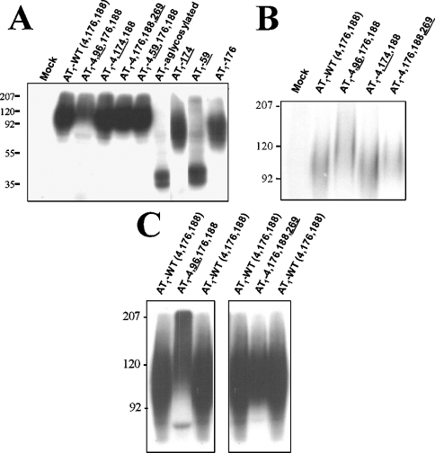
Figure 2. Photoaffinity labelling of AT1 receptor glycosylation mutants.
(A) COS-7 cells expressing AT1-WT and various mutant receptors were incubated in the presence of 5 nM 125I-[L-Bpa8]AngII for 1 h at room temperature. The cells were irradiated under 365 nm filtered UV light for 45 min at 0 °C. After solubilization, the samples were resolved on 10% SDS/PAGE followed by autoradiography, as described in the Experimental section. Electrophoresis of selected mutant receptors was prolonged for 2 h with a small (B) and large (C) amount of specifically photolabelled receptor complex to highlight the shift in electrophoretic mobility. Protein standards of the indicated molecular-mass markers were run in parallel (values shown on the left-hand side of the gels in kDa). These results are representative of at least three independent experiments.

